Sponsored by the UCLA Brain Mapping Center Faculty
The focus of these talks is on advancing the use of brain mapping methods in neuroscience with an emphasis on contemporary issues of neuroplasticity, neurodevelopment, and biomarker development in neuropsychiatric disease.
Hosted By: Shantanu Joshi, PhD, Neurology, UCLA
 |
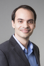
Justin Haldar, PhD Associate Professor Electrical and Computer Engineering and Biomedical Engineering, USC |
Magnetic resonance (MR) imaging technologies provide unique capabilities to probe the mysteries of biological systems, and have enabled novel insights into anatomy, metabolism, and physiology in both health and disease. However, while MR imaging is decades old and has already revolutionized fields like medicine and neuroscience, current methods are still far from fully delivering on the potential of the MR signal. In particular, traditional methods are based on classical sampling theory, and suffer from fundamental trade-offs between signal-to-noise ratio, spatial resolution, and data acquisition speed. These issues are exacerbated in high-dimensional applications, due to the curse of dimensionality. Our work addresses the limitations of traditional MR imaging using signal processing approaches that have recently become practical because of improvements in modern computational capabilities. These approaches are possible because of certain "blessings of dimensionality," e.g., that high-dimensional data often possesses unexpectedly simple structure which can be exploited to alleviate the classical barriers to fast high-resolution imaging. This seminar will describe approaches we have developed that use novel constrained imaging models (based on sparsity, partial separability, linear predictability, etc.) to guide the design of new MR data acquisition and image reconstruction methods, and enable substantial acceleration of both low-dimensional and high-dimensional MR imaging experiments. These methods will be illustrated in the context of applications such as fast high-resolution T1-weighted anatomical imaging, fast sub-millimeter diffusion imaging, ungated free-breathing cardiac imaging, and novel high-dimensional diffusion-relaxation hybrid experiments that provide unique insights into tissue microstructure.